Analysis of Fluorescence Polarization competition assays with AFFINImeter

Fluorescence Polarization competition assays are widely used in numerous research fields to determine the affinity of unlabeled ligands that compete with a fluorescent probe for binding with the same macromolecule. Herein, we show how to perform a thoughtful analysis of these experiments with AFFINImeter for spectroscopic techniques, to elucidate, not just IC50 values, but also a quantitative direct measurement (KA) of the binding affinity.


Introduction


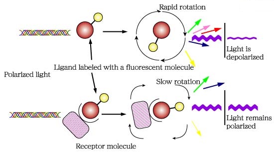
Fluorescence polarization (FP) is a powerful technique that nowadays is widely utilized in high-throughput screening (HTS) and drug discovery (1). In FP assays monitoring a binding event is possible mainly because this technique is sensitive to changes in molecular weight. The assay requires of a fluorescent molecule (the probe) that is excited by plane-polarized light. For small molecules, the initial polarization decreases rapidly due to rotational diffusion during the lifetime of the fluorescence, which results in low fluorescence polarization. When the small fluorescent molecule binds to a larger molecule (and consequently of slower rotation) an increased fluorescent polarization is observed (Fig. 1).
Advances in experimental aspects of the technique like assay design and fluorescent probes is enabling the application of FP to increasingly complex biological processes (1).

Fig.1. The principle of fluorescence polarization (2).


Frequently, a competition displacement assay format is used in FP experiments where a fluorescent labelled molecule bound to a macromolecule is displaced by an unlabeled molecule with the consequent decrease of polarization. This assay yields a quantitative measure of IC50 values (half maximal inhibitory concentration) of the unlabeled competitors. However, IC50 does not provide a direct measure of affinity and the calculation of the binding constant (KA) is often desirable. In this sense, the software AFFINImeter Spectroscopy offers exclusive analysis tools to perform a thoughtful analysis of FP competition assays, to directly deliver values of binding constants of the interaction between the competitor and the macromolecule. As an illustrative example, we present herein the use of AFFINImeter Spectroscopy for the analysis of FP competition assays to characterize oligosaccharide –protein interactions.


FP competition assays to study the binding between midkine and chondroitin sulfate-like tetrasaccharides


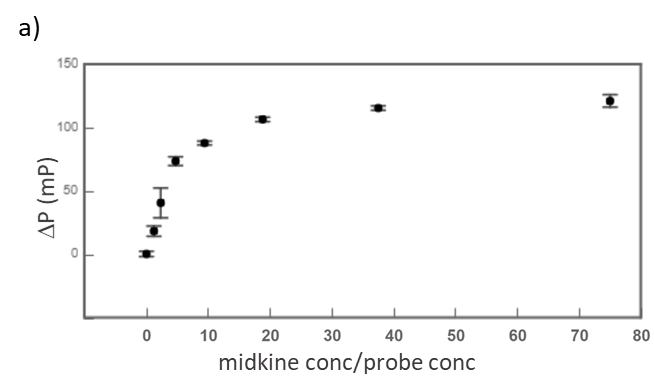
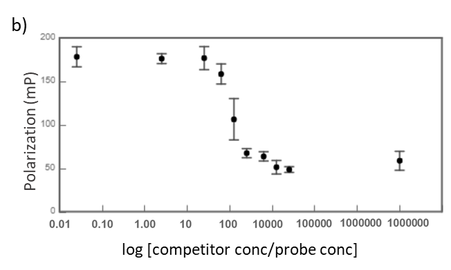
Midkine is a cytokine which biological activity is regulated by its binding with glycosaminoglycans (GAGs), such as heparin and chondroitin sulfate. To better understand these recognition processes Pedro M. Nieto et al. have recently reported the binding of a series of synthetic chondroitin sulfate-like tetrasaccharides with midkine, using FP competition assays (3). First, the direct binding of midkine and a fluorescein labelled heparin hexasaccharide (fluorescent probe) was monitored in a direct FP titration (Fig. 2a). Second, FP competition assays were performed, in which the polarization of samples containing fixed concentrations of midkine and fluorescent probe was recorded in the presence of increasing concentrations of different synthetic sugars to obtain the corresponding IC50 values (Fig. 2b) (3).


Fig.2. a) Binding curve of the titration of fluorescent probe with midkine 3a;b) Representative competition curve (semi-log plot) of an FP competition assay where the fluorescent probe is displaced from a pre-formed complex with midkine by the competitor.


Analysis of FP competition assays with AFFINImeter Spectroscopy


The determination of KA of the interaction of the unlabeled ligand with the macromolecule in an FP competition assay is possible if the corresponding curve is analyzed using a competitive binding model where two equilibria are described, one between the free species and the macromolecule–probe complex and one between the free species and the macromolecule–competitor complex. In this analysis, the KA value of the interaction between probe and macromolecule can be fixed (as it can be previously calculated from the direct binding experiment) in order to determine KA of the competitor-macromolecule complex. Yet, a more robust approach consists of a global analysis of direct and competitive curves where constraints between experimental parameters are imposed. Using this approach, we have used AFFINImeter Spectroscopy in the analysis of data from the direct measurement of the fluorescent labelled hexasaccharide binding to midkine and together with data from the displacement assay using an unlabeled, synthetic disaccharide as a competitor (4).

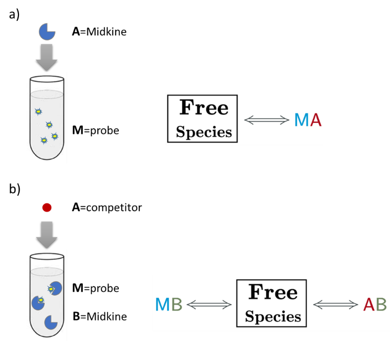
An AFFINImeter fitting project was generated where the curve from the direct titration and two curves from replicates of the competitive assay were included. A 1:1 simple binding model and a competitive model were used to fit direct and competitive data, respectively (Fig. 3).

Fig.3. Schematic representation of the binding models used to fit the FP data.5 These models can be easily built with the “model builder” tool of AFFINImeter where a) in the 1:1 model “M” represents the species sensitive to binding (fluorescent probe) and “A” represents the titrant (midkine); b) in the competitive model “M” is the sensitive species (probe), A is the titrant (competitor) and B is a third species involved in the event (midkine in a preformed complex with the probe).


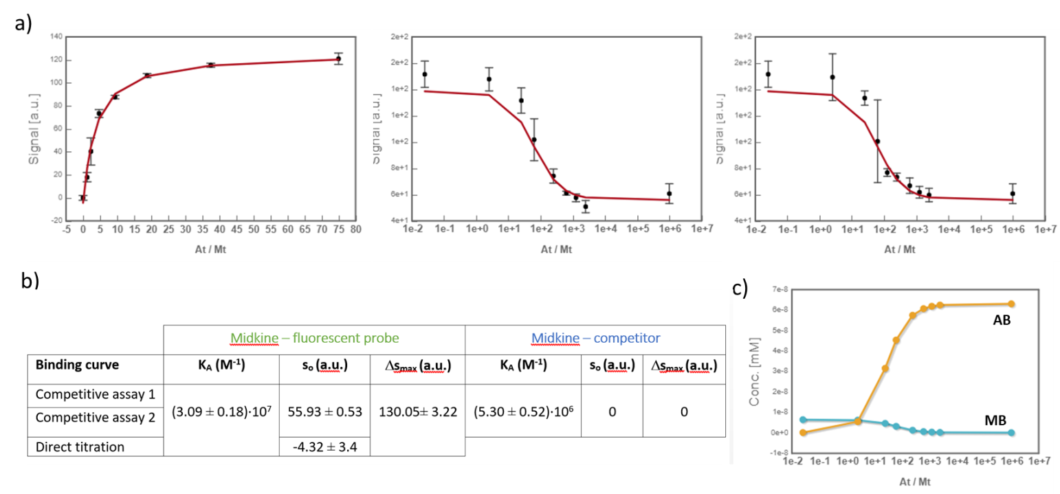
In the analysis, the fitting parameters considered were the KA of each complex, the signal value of the unbound state (s0), and the maximum signal change (Δsmax) of the midkine-probe complex formation. Δsmax of the midkine-competitor complex is equal to zero because the competitor is not fluorescent. Since midkine-probe binding is present in both models, restrictions were made considering that KA and Δsmax of the 1:1 model (FS↔MA) are equal to the same parameters describing this equilibrium in the competitive model (FS↔MB). Besides, s0 was common between replicates of the competitive assay. It is worth to mention that the curves analyzed are the mean of three replicate experiments and the corresponding standard deviation of the curve data points are also considered in the fitting process.  The global analysis performed in this way returned the KA of the interaction of midkine with the probe, (3.09 ± 0.18)*107 M-1, and with the competitor, (5.30 ± 0.52)*106 M-1 (Fig. 4). Additionally, AFFINImeter automatically generates the species distribution plot, valuable in the interpretation of results. The species distribution plot of Fig. 4c shows the displacement of the probe by the unlabeled disaccharide in the competition assay.


Fig.4. a) Global analysis of FP curves from the direct titration of probe (10 nM) with midkine (12 – 750 nM) and from a competitive assay consisting of a sample with probe (10 nM) and midkine (63 nM) where the competitor is added at increasing concentrations (0 – 20 mM). Curves from two replicates of the competitive assay were used in the analysis. All the polarization values are the average of three replicate wells and the error bars represent the corresponding standard deviation; b) values of KA, s0 and Δsmax determined for each equilibrium; c) species distribution semi-log plot of the competitive assay.


Conclusions


This case study exemplifies the utility of AFFINImeter Spectroscopy in the analysis of FP competitive binding assays. The advantages of using this software rely on the possibility to obtain reliable KA values of the binding between competitor and macromolecule from a global analysis where KA and Δsmax of the probe-macromolecule complex, are shared parameters between curves (they are not pre-determined fixed parameters). Besides, standard deviation between replicates is taken into account in the fitting process. Ultimately, these tools provide a more robust analysis and reliable characterization of binding interactions monitored through competitive assays.


Try AFFINImeter Spectroscopy

Acknowledgements


We would like to thank Dr Pedro Nieto Mesa and Dr José Luis de Paz Carrera from the Institute of Chemical Research (IIQ) of the Spanish National Research Council (CSIC), for kindly share with us the FP data presented herein.


References & Notes


1 Hall, M.; Yasgar, A.; Peryea, T.; Braisted, J.; Jadhav, A.; Simeonov, A.; Coussens, N. Fluorescence Polarization Assays In High-Throughput Screening And Drug Discovery: A Review. Methods and Applications in Fluorescence 2016, 4, 022001.

2 This figure has been taken from http://glycoforum.gr.jp/science/word/glycotechnology/GT-C06E.html.

3 a) Solera, C.; Macchione, G.; Maza, S.; Kayser, M.; Corzana, F.; de Paz, J.; Nieto, P. Chondroitin Sulfate Tetrasaccharides: Synthesis, Three-Dimensional Structure And Interaction With Midkine. Chemistry – A European Journal 2016, 22, 2356-2369; b) Maza, S.; Gandia-Aguado, N.; de Paz, J.; Nieto, P. Fluorous-Tag Assisted Synthesis Of A Glycosaminoglycan Mimetic Tetrasaccharide As A High-Affinity FGF-2 And Midkine Ligand. Bioorganic & Medicinal Chemistry 2018, 26, 1076-1085.

4 The competitor is a synthetic disulfated disaccharide described in ref. 3b as compound 18.

5 These model were created with the “model builder” tool, exclusive of AFFINImeter. For more information go to AFFINImeter knowledge center.
Working with AFFINImeter models based on a stoichiometric equilibria approach

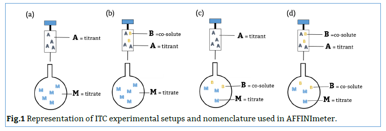
The appropriate design and use of binding models in AFFINImeter pass through an understanding of the nomenclature that the software uses to describe a given experimental setup and the species that take part in the assay.
AFFINImeter contemplates the presence of up to three species participating in the experiment: 1) the titrant, or compound placed in the syringe; 2) the titrate, or compound in the calorimetric cell and 3) a co-solute or third compound that can be in the syringe and/or in the cell. These species are labelled in the reaction builder as follows:

As illustrated in Fig. 1, M always refers to the compound placed in the calorimetric cell and A always refers to the compound in the syringe. B always refers to a third component that can be in the syringe (Fig. 1b), in the cell (Fig. 1c) or in both places at once (Fig. 1d):


Once the nomenclature of  AFFINImeter is clear, you can download this note that our scientific team have prepared to understand how our users can be working with models based on a stoichiometric equilibria approach:

Download

You can also check our latest post where we describe “The concepts of stoichiometric and site binding constants”


Pursuing a reliable calculation of binding affinity.

The relevance of uncertainties.


The reliable determination of binding constants (Ka) and enthalpies (ΔH) from Isothermal Titration Calorimetry depends, among other aspects, on the quality of the thermogram recorded where a high signal-to-noise ratio is pursued to get a trusty isotherm. Low signal-to-noise ratio conducts to less precise integration of the thermogram peaks and, consequently, to a binding isotherm defined by points with larger related uncertainties. Logically, the uncertainty associated to the integral points will significantly affect the determination of the thermodynamic parameters and should not be set aside through the data analysis.

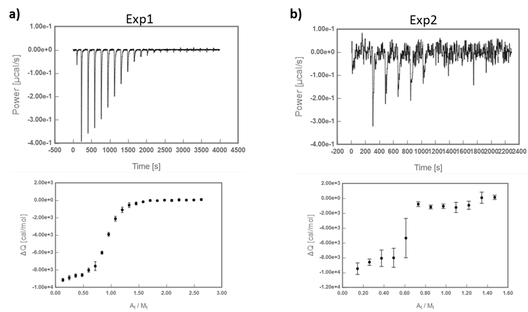
Here´s an illustrative example of two ITC experiments of the interaction between Carbonic Anhydrase II (CA) and 4-carboxybenzenesulfonamide (4-CBS) that yielded thermograms of different quality (Fig 1*).

Fig.1 ITC titrations of 4-CBS into CA. a) Exp1, higher signal-to-noise ratio; b) Exp2, lower signal-to-noise ratio.

Data fitting to a 1:1 binding model was performed for both experiments and for comparison purposes the analyses were conducted twice, taking into account the uncertainties associated to the calculation of the peaks integral (weighted fitting) and not considering them (all the points having the same weight).
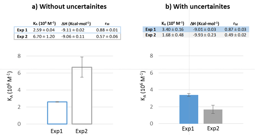
The results show a larger discrepancy in the Ka values obtained from Exp2, depending on whether or not the uncertainties are included in the fitting. Besides, the Ka obtained from the weighted fitting of Exp2 is significantly closer to the corresponding Ka value of the weighted fitting of Exp1 (Figure 2).
Fig.2 KA, DH and rM (correction parameter for CA concentration in the cell) values from fitting of Exp1 and Exp2 a) without uncertainties and b) with uncertainties. The bar diagrams show the comparison between KA values of Exp1 and Exp2.


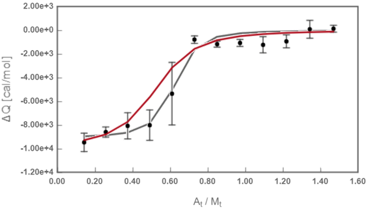
The main origin of this difference is the large uncertainty associated to the mid-titration point, which consideration has an effect on the final Ka value (Figure 3).
Fig.3 Fitted isotherm from Exp 2. Redline: theoretical curve calculated when considering uncertainties; black line: theoretical curve calculated with no uncertainties.


Being aware of the relevance of weighting data points based on their uncertainty AFFINImeter includes, into the raw data processing step, the automatic determination of uncertainties in the peaks integration and the possibility to perform weighted fitting for a more reliable analysis. Another good reason to work with AFFINImeter!


Solving complex interactions with AFFINInimeter: Competing ligands binding to a multiple site receptor

Isothermal Titration Calorimetry (ITC) is a versatile technique with the potential of deconvoluting the various binding events that may coexist in complex interactions. In this sense, a major drawback has been the lack of mathematical models and computational tools to properly analyze such experiments. AFFINImeter counts with an advanced functionality, the “Model Builder” with which researchers can easily design their own binding models through the combination of distinct (coupled) binding equilibria, to obtain thermodynamic and structural information from complex ITC experiments.

Two Competing ligands binding to a receptor with two sites

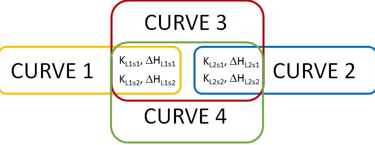
As a demonstration of the potential of AFFINImeter and its “Model Builder”, the analysis of an ITC experiment involving a receptor “M” (biomacromolecule) with two different binding sites (s1 and s2) that may accommodate two competing ligands (“A” and ”B”) is presented here.

Download complete case here: Competitive Binding case of use

Expanding the range of applications of ITC in the Pharmaceutical Industry with AFFINImeter: A practical Case.

Many Drug–receptor interactions are characterized by complex binding modes that are far away from the behavior of a standard 1:1 model. This is the case of Heparin (Hp), one of the most commonly prescribed anticoagulant drugs, which exerts its effect through its interaction with the serine protease Antithrombin (AT-III). Hp is a linear heterogeneous polysaccharide containing a specific pentasaccharide sequence that binds AT-III with high nanomolar affinity (responsible for the anticoagulant activity); but AT-III also binds other Hp sequences with lower affinity. Determining the content of AT-III binding pentasacchride in Low Molecular Weight (LMW) Heparins is a requirement for Pharmaceutical companies that manufacture this type of anticoagulants; due to the intrinsic heterogeneity of Hp, obtaining this information it is not straightforward (1).

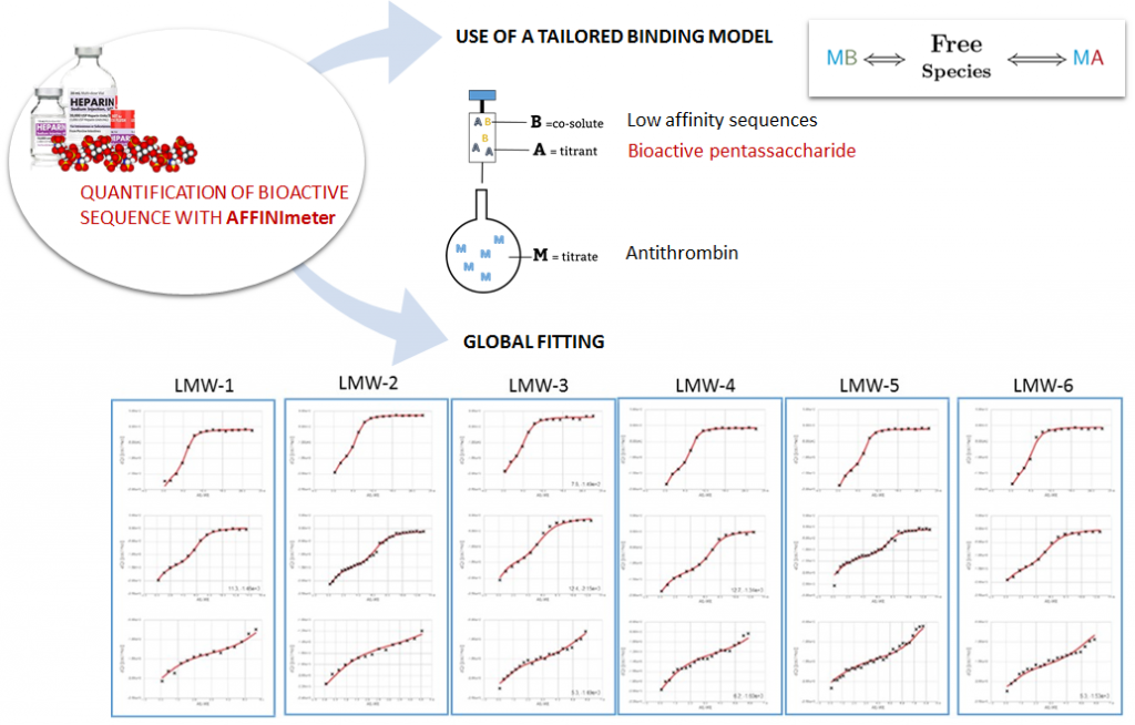
We have developed a new protocol based on ITC and AFFINImeter to determine the content of AT-III binding pentasaccharide in Heparins, which is summarized in the following scheme:

New method based on AFFINImeter to determine the content of AT-III binding pentasacchride in LMW Hp: 1) use of a tailored binding model that describes the competitive binding between the pentasaccharide (A) and other low affinity sequences (B) with AT-III (M); 2) global fitting of several isotherms registered under different Hp and or AT-III concentrations where the parameters rA and rB (that account for the fraction of A and B in the Hp sample) are fitting parameters and common among the different isotherms.


This method illustrates the great potential of the model builder and global fitting AFFINImeter tools to develop protocols of practical utility in the Pharmaceutical industry (2). We have successfully validated the protocol in the analysis of unfractionated Hp and a series LMW Hp in collaboration with the Pharmaceutical company Laboratorios Rovi (http://www.rovi.es/).

References

  1. Nandurkar H., Chong B, Salem H, Gallus A, Ferro V, McKinnon R. Low-molecular-weight heparin biosimilars: potential implications for clinical practice. Internal Medicine Journal, 2012, 44(5), pp 497–500.

  2. For a detailed description of the protocol contact us at support@affinimeter.com

Global fitting analysis of a protein-ligand binding experiment

Global fitting analysis of a protein-ligand binding experiment

A few weeks ago AFFINImeter launched an Isothermal Titration data Analysis challenge of the analysis of a protein-ligand binding experiments. The participant had to globally analyze a set of Isothermal Titration Calorimetry experiments using AFFINImeter and get the thermodynamic and structural parameters of the interaction between both molecules (the receptor protein and the ligand).

The participants in this contest had the opportunity to demonstrate their ability to propose the right model for a given binding isotherm as well as to get the corresponding parameters upon fitting using AFFINImeter. On their side, less experienced participants had the opportunity to:

The results recently published by Henlz et al in [Methods 59 (2013) 336-348] were taken as a reference to generate the set of ITC isotherms selected for this contest.

(more…)

How to perform a Global Fitting Analysis?

The Global fitting of multiple isotherms is one of the advanced tools that AFFINImeter offers to facilitate the analysis and interpretation of isothermal titration experiments and to expand the range of applications of this technique.

The following video tutorial describes the global fitting of three isotherms of a displacement assay describing, a receptor interacting with a tight ligand, with a weak ligand, or with both ligands simultaneously, in a competitive experiment where the ligands are mixed in the syringe of the ITC equipment.


If you want to know more about global fittings with AFFINImeter you can also download the case of use “Global Analysis in ITC Displacement Titrations with AFFINImeter” that describes a Displacement Titration Assay to determine the thermodynamics of HIV-protease with indinavir, a high-affinity binder, and with acetyl-pepstatin, a weaker ligand.

ITC displacement titrations offer an attractive alternative to standard assays when working with ultra-high or ultra-low- affinity interacting systems. The method requires the fitting of at least two isotherms that share various adjustable parameters. The case study exemplifies the potential advantages of using AFFINImeter in ITC displacement assays. The software offers unique advanced tools that enhance the robustness of the method and makes it more versatile, facilitating the acquisition of reliable thermodynamic data from ultra-high of ultra-low affinity systems. Thus, it opens a door for new applications of the displacement assay.


Global Fitting Analysis in ITC Displacement Titrations with AFFINImeter

ITC displacement titrations offer an attractive alternative to standard assays when working with ultra high- or ultra low- affinity interacting systems. The method requires the fitting of at least two isotherms that share various adjustable parameters. AFFINImeter counts with advanced tools, like the global fitting of multiple dataseries and the analysis of isotherms registered under unusual experimental design, which can facilitate the analysis and expand the range of applications of isothermal titration calorimetry experiments. As an illustration, herein we present a displacement titration assay to determine the thermodynamics of HIV-protease with indinavir, a high affinity binder, and with acetyl-pepstatin, a weaker ligand. Using AFFINImeter a global analysis of four isotherms was performed describing: HIV-protease binding to indinavir (I) or to acetyl-pepstatin (II): HIV-protease binding to indinavir incorporating acetyl-pepstatin in the cell (III) or in the syringe (IV).

Isothermal Titration Calorimetry is one of the most commonly used approaches to obtain affinity and thermodynamic data of molecular interactions and has become a routine method in the pharmaceutical industry.1 Isothermal titration Calorimetry is applicable to numerous interacting systems, as long as a detectable heat change is produced during complexation, covering an important range of binding affinities (106 ≤ KA ≤108 M-1). Nevertheless, standard ITC experiments present some limitations in the case of very low- or very high-affinity interactions (i.e. affinities in the low millimolar or high nanomolar range, respectively). High affinity interactions (KA ≥ 109 M-1) yield square-shaped isotherms whose fitting yield accurate values of the binding enthalpy but only estimates of the association constant. Attempts to recover a sigmoidal shape requires the use of very low concentrations of the interactants that, in most cases, is not feasible in the practice (the minimum concentration that will typically cause a confidently measurable heat change for a 1:1 interaction is about 10 μM). On the opposite, low affinity interactions should be studied at high concentrations and this requirements is often a serious limiting step due to various potential reasons like limited solubility and/or availability of the sample molecule, or the existence of aggregation processes at the required concentration. In both high- and low affinity systems these experimental drawbacks can be circumvented by using the ITC displacement method.2,3 Here, the receptor is titrated with a high affinity ligand, but in the presence of a weaker ligand in the sample cell that competes for the complexation with the receptor (figure 1). With this experimental set up the apparent affinity of the strong ligand is “artificially” lowered, obtaining a sigmoidal isotherm that yields more accurate binding data. When the goal is to obtain the thermodynamic parameters of an ultra highaffinity system, a titration with the weaker binder is performed first to obtain the corresponding affinity constant and enthalpy (KA-weak and H-weak). These values are required for the analysis of the ITC displacement experiment, where a competitive binding model is used to estimate the thermodynamic parameters of the tight binding (KA-tight and H-tight). Analogously, when the goal is to obtain information of an ultra low- affinity system a direct ITC titration of the receptor alone with a ligand of higher affinity is performed. The resulting KA-tight and H-tight are then incorporated in the analysis of the isotherm from the ITC displacement assay.

This case study exemplifies the potential advantages of using AFFINImeter in ITC displacement assays. The software offers unique advanced tools that enhance the robustness of the method and makes it more versatile, facilitating the acquisition of reliable thermodynamic data from ultra-high of ultra-low affinity systems. Thus, it opens a door for new applications of the displacement assay.
1 G. Holdgate, S. Geschwindner, A. Breeze, G. Davies, N. Colclough, D. Temesi, L. Ward, Biophysical Methods in Drug Discovery from Small Molecule to Pharmaceutical. Protein-Ligand Interactions. In Methods in Molecular Biology 2013, 1008, pp 327-355.
2 A. Vellazquez-Campoy and E. Freire, Isothermal titration calorimetry to determine association constants for highaffinity ligands. Nature protocols 2006, 1, pp 186-191.
3 W. B. Turnbull, Divided we fall? Studying low-affinity fragments of ligands by ITC. GE Healthcare Life Sciences protocol.


Download the case of use here

Isothermal Titration Calorimetry
Global Analysis in ITC Displacement Titrations with AFFINImeter